Nasal Cavity Anatomy Ct : Paranasal Sinuses Ct Anatomy W Radiology : The ct test is usually made to evaluate the anatomy of the paranasal sinuses.
Get link
Facebook
X
Pinterest
Email
Other Apps
Nasal Cavity Anatomy Ct : Paranasal Sinuses Ct Anatomy W Radiology : The ct test is usually made to evaluate the anatomy of the paranasal sinuses.. *they are separated from each other by a septum. Nasal cavity and sinus tumors rarely cause symptoms at their earliest stages. The nasal cavity opens into a network of sinuses: Cribriform plate of the ethmoid. Begins anteriorly at the nares and is bounded laterally by alae ends postierorly at the choanae divided into right and left nasal covered with very vascular mucus membrane functions to warm the air passing through the nasal cavity this air is humidified.
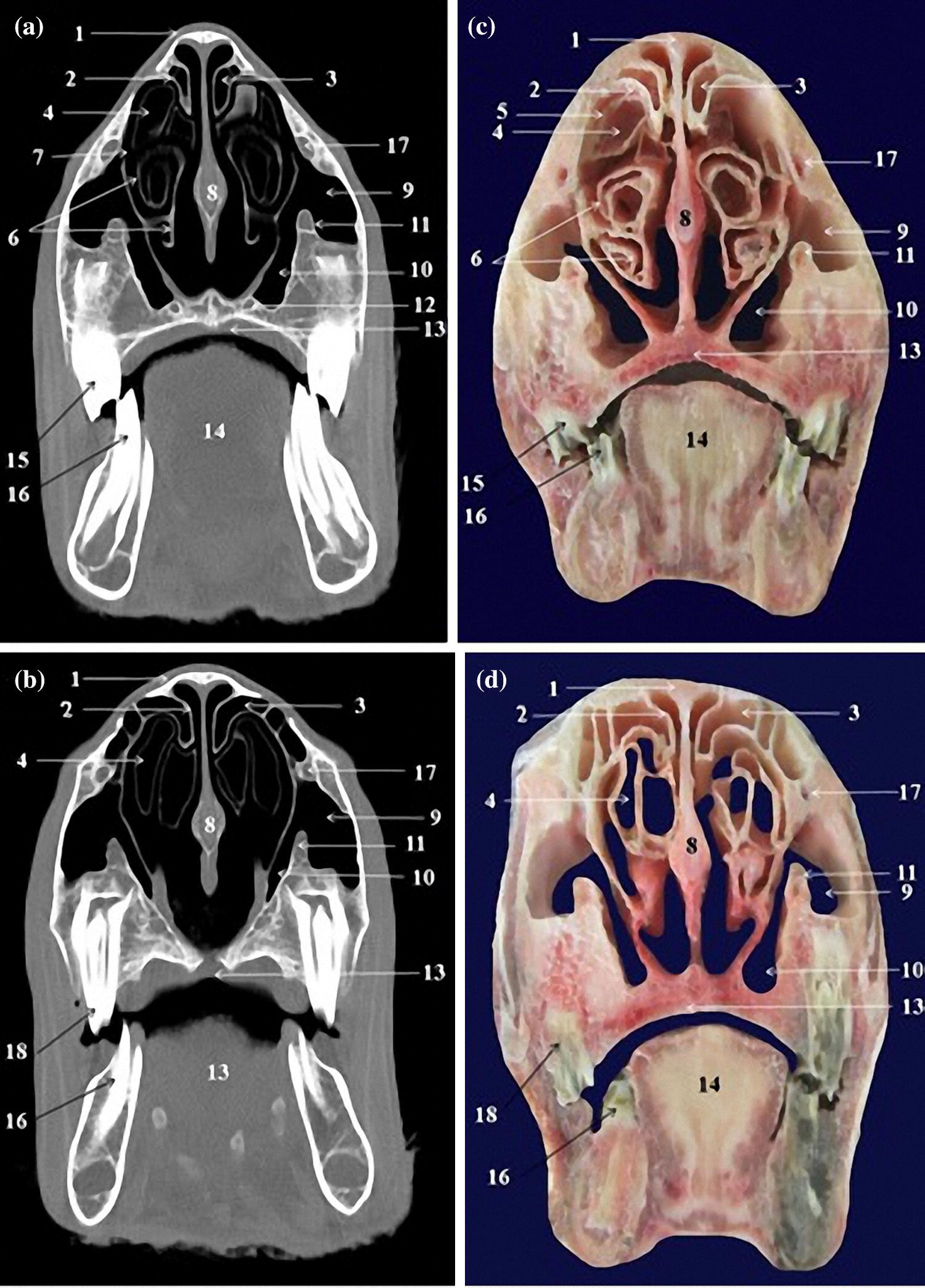
Knowledge of nasal cavity anatomy facilitates comprehension of the pattern of spread of tumors of nasal cavity carcinomas spread to adjacent sinuses depending on the location of origin: The framework of the nose consists of bone and cartilage. Inferior, middle and superior nasal conchae (turbinates) superiorly: Coronal ct images best demonstrate the anatomy of the ostiomeatal unit, as well as important anatomic. Nasal cavities are located in the midface, separated by a median septum;
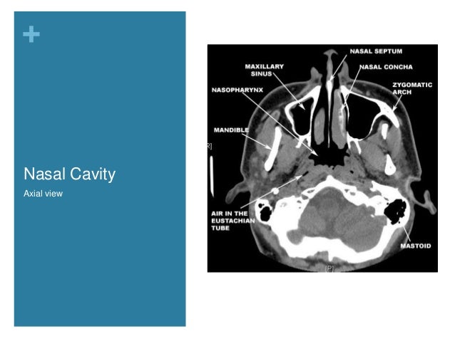
The Preoperative Sinus Ct Avoiding A Close Call With Surgical Complications Radiology from pubs.rsna.org But did you know that 80% of taste actually comes from what we smell? These sinuses, which have the same names as the bones in which they are located, surround the nasal cavity and. Overview of nasal cavity and its boundaries. Ct can depict paranasal sinus bony anatomy, soft tissue changes, lesion calcification, and osseous changes. Brain, bones of skull, paranasal sinuses. Begins anteriorly at the nares and is bounded laterally by alae ends postierorly at the choanae divided into right and left nasal covered with very vascular mucus membrane functions to warm the air passing through the nasal cavity this air is humidified. Is your nose also an excretory organ? Gross anatomy the nasal cavity is formed by 1:
The nasal septum divides the cavity into two cavities, also known as fossae.
Is it nasal cavity or cavities? Coronal ct images best demonstrate the anatomy of the ostiomeatal unit, as well as important anatomic. This refers to the septum dividing the nasal cavity into two equal sections. The nasal anatomy shows much individual variation. In this article, we shall look at the applied anatomy of the nasal cavity, and some of the relevant clinical syndromes. Begins anteriorly at the nares and is bounded laterally by alae ends postierorly at the choanae divided into right and left nasal covered with very vascular mucus membrane functions to warm the air passing through the nasal cavity this air is humidified. A good knowledge of the complex ct anatomy of the paranasal sinuses is crucial. Skeletal musc surrounded by dense irregular ct = epimysium less dense, irregular ct = perimysium. 3 name the structures opening into the lateral wall of nasal cavity. Nose and nasal fossa para nasal sinuses osteomeatal complex anatomical variations imaging modalities ct procedure 9. Ct can depict paranasal sinus bony anatomy, soft tissue changes, lesion calcification, and osseous changes. Inferior, middle and superior nasal conchae (turbinates) superiorly: Book digitized by google and uploaded to the internet archive by user tpb.
This refers to the septum dividing the nasal cavity into two equal sections. Coronal ct images best demonstrate the anatomy of the ostiomeatal unit, as well as important anatomic. The nasal anatomy shows much individual variation. Gross anatomy the nasal cavity is formed by 1: This is the site where the artery is most liable to injury.
Figure 3 Anatomy Of The Head In The Saanen Goat A Computed Tomographic And Cross Sectional Approach Springerlink from media.springernature.com Allergic polyps are usually bright red because of their extensive network of blood vessels. Is it nasal cavity or cavities? Brain, bones of skull, paranasal sinuses. Check out this ultimate guide to studying anatomy. A anterior nasal fossa, level of the uppermost insertion of both uncinate processes on the. The nasal cavity via the cribriform plate. • each nasal cavity has a floor, roof, medial wall nerves of nasal cavity: The nasal cavity also contains structures to detect chemical odorants and resonate the voice.
Knowledge of nasal cavity anatomy facilitates comprehension of the pattern of spread of tumors of nasal cavity carcinomas spread to adjacent sinuses depending on the location of origin:
• each nasal cavity has a floor, roof, medial wall nerves of nasal cavity: Knowledge of nasal cavity anatomy facilitates comprehension of the pattern of spread of tumors of nasal cavity carcinomas spread to adjacent sinuses depending on the location of origin: *they are separated from each other by a septum. Allergic polyps are usually bright red because of their extensive network of blood vessels. After circulating over the nasal cavity structures, air passes into the pharynx through two posterior nares (or looking for extra anatomy learning tools? Overview of nasal cavity and its boundaries. Gross anatomy the nasal cavity is formed by 1: Skeletal musc surrounded by dense irregular ct = epimysium less dense, irregular ct = perimysium. Maxillary sinuses are in the cheek area, below the eyes on either side of the nose. Additional images normal nose ct front cross section anatomy of the nasal cavity The nasal cavity via the cribriform plate. Each cavity is the continuation of one of the two nostrils. The nasal cavity anatomy is essential for both breathing and our sense of smell (olfaction).
In this page, we are going to study the nose anatomy, with a special focus on the anatomical importance of the nasal cavity structure. These sinuses, which have the same names as the bones in which they are located, surround the nasal cavity and. Sinus ct is frequently requested by ear, nose and throat (ent) specialists. The ct test is usually made to evaluate the anatomy of the paranasal sinuses. The nasal cavity opens into a network of sinuses:
Nasal Cavity And Paranasal Sinuses Radiologic Anatomy from image.slidesharecdn.com Dural venous sinuses, veins, arteries. Maxillary sinuses are in the cheek area, below the eyes on either side of the nose. Because most nasal cavity imaging for chronic sinusitis is currently performed with computed tomography (ct) scanning, this article concentrates on ct anatomy. …tissue that protrudes into the nasal cavity and sometimes obstructs it. Cribriform plate of the ethmoid. 4 describe the arterial supply of nasal septum. Ct scanning is painless, noninvasive and accurate. Ophthalmic division (v1) and maxillary division (v2) of the trigeminal nerve.
In this page, we are going to study the nose anatomy, with a special focus on the anatomical importance of the nasal cavity structure.
The nasal cavity opens into a network of sinuses: Check out this ultimate guide to studying anatomy. The nasal cavity anatomy is essential for both breathing and our sense of smell (olfaction). Brain, bones of skull, paranasal sinuses. …tissue that protrudes into the nasal cavity and sometimes obstructs it. But did you know that 80% of taste actually comes from what we smell? • separated by a midline nasal septum. This is the site where the artery is most liable to injury. Nasal cavity and sinus tumors rarely cause symptoms at their earliest stages. Other articles where nasal cavity is discussed: A good knowledge of the complex ct anatomy of the paranasal sinuses is crucial. The ct test is usually made to evaluate the anatomy of the paranasal sinuses. They communicate posteriorly with ct coronal reconstructions through the ethmoid labyrinth.
Book digitized by google and uploaded to the internet archive by user tpb nasal cavity anatomy. Brain, bones of skull, paranasal sinuses.

Comments
Post a Comment